Patient Education
Everything you could want to know about common eye problems. Learn what to look for, what you can do, and how we can help you avoid and treat the symptoms. Please Contact Us with any further questions or concerns about your eyes.
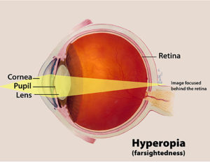
Farsightedness or hyperopia occurs when the eyeball is too short for the focusing power of the lens and cornea. This causes light rays to focus behind the retina. As a result, the eye sees distant objects more clearly while near objects appear blurred. In essence, the eye is underpowered. To correct hyperopia, a “plus” lens containing additional optical power is needed to permit sharp vision of near objects.
The shape of a hyperopic eye focuses images behind the retina, resulting in blurred near vision. A ‘plus’ lens increases the focusing power of the eye correctly focusing the image on the retina producing clear vision at all distances.
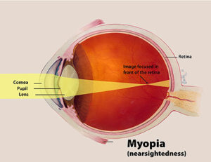
Nearsightedness or Myopia occurs when the eyeball is too long for the focusing power of the lens and cornea. The result is an overpowered eye that causes images to focus in front of the retina. A myopic eye sees near objects within a certain range very clearly while distance vision appears blurry at all times. An estimated 70 million people in the U.S. suffer from myopia. To correct myopia, a ‘minus’ lens is required to push the image to the back to the retina permitting sharp distance vision.
The shape of a myopic eye focuses the image in front of the retina resulting in blurred distance vision. A ‘minus’ lens decreases the focusing power of the eye correctly focusing the image on the retina producing clear vision at all distances.
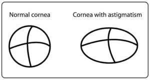
Astigmatism is a common visual distortion caused by an irregularly shaped cornea. The surface of the cornea is toric, oblong in shape like the side of a football, instead of perfectly spherical like a basketball. One surface or axis of the football is long with a shallow curve while the other surface is short with a steep curve. Light rays passing through an oblong cornea bend unequally, causing two focusing points. The result is blurred vision at most distances.
Astigmatism is typically present at birth. Over time the condition may slowly increase but generally it remains relatively stable over a lifetime. Forty-five percent of people who require vision correction have some degree of astigmatism. Symptoms include squinting, occasional headaches and eye strain. Astigmatism often accompanies myopia and hyperopia.
The irregular shape of an astigmatic eye produces two focusing points around the retina. The result is blur at all distances. A ‘toric’ lens corrects each toric irregularity so that light is properly focused on the retina producing clear vision at all distances.
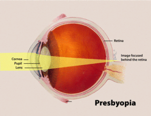
Presbyopia, Greek for “aging eye,” is caused by a combination of natural aging, the hardening of the eye’s crystalline lens, a transparent body in the front of the eye that serves to focus light rays on the retina, and a weakening of the focusing muscles. As people reach their 40s, the crystalline lens grows thicker and begins to lose its elasticity. Gradually, the eye muscle control diminishes and people find it increasingly difficult to focus on near objects. To compensate for the reduced focusing ability the tendency is to hold reading material further away. Eventually the arms become too short resulting in blurred vision and eye strain.
To correct this condition lenses of different powers are required. The first lens is a lens to correct the distance vision and the second lens provides the focusing power for reading. This is easily seen in bifocal spectacles. The most common consequence of bifocals is the loss clarity for intermediate distances including computer work. Unfortunately, each lens strength has a very specific distance from the nose in which things will be clear. The same lens needed for reading will NOT work for seeing something a little further away such as the computer. To see the distance needed for the computer, a different strength is needed but that strength will not work for reading. Ultimately, to adequately function in life different lens strengths are needed for every distance. The ideal lens for correcting presbyopia is the progressive lens. This is a molded lens that provides power for all distances including the intermediate distances. Additionally this is a much more natural lens for your brain to accept. Where the bifocal only allows clear vision for two distances, the progressive simulates the eyes natural ability to have clear vision for all distances.
Dry eye syndrome is a chronic lack of sufficient lubrication and moisture in the eye. Consequences extend from subtle but constant irritation to ocular inflammation of the front of the eye. Dry eye will often result in stinging, burning, scratchy sensation, sensitivity to light, tearing, tired eyes, and difficulty wearing contact lenses, as well as blurred or fluctuating vision, often worsening at the end of the day or after visually focusing for a prolonged period on a nearby task. It is particularly bothersome for wearers of contact lenses, the elderly, certain lifestyles, medications, and visual demands.
Signs of Dry Eye
Persistent dryness, scratching and burning in your eyes are signs of dry eye syndrome. Some people also experience a “foreign body sensation,” the feeling like there’s something in the eye. Occasionally watery eyes can result from dry eye syndrome, because the excessive dryness works to over stimulate the watery component of your eye’s tears. Sometimes the eye doesn’t produce enough tears, or the chemical composition of the tears causes them to evaporate too quickly. Other times dry eye results when you don’t blink often enough. Computer users, for example, often forget to blink for long periods of time, or the monitor is too high so even if their tear film is normal, it eventually evaporates, leading to discomfort.
Causes of Dry Eye
Dry eye syndrome has several causes. It occurs as a part of the natural aging process, especially during menopause; as a side effect of such medications as antihistamines, antidepressants and birth control pills; the use of cigarettes, marijuana, or because you live in a dry, dusty or windy climate. Dry eyes are also a symptom of systemic diseases such as lupus, rheumatoid arthritis or Sjogren’s syndrome (a triad of dry eyes, dry mouth, and rheumatoid arthritis or lupus). Long-term contact lens wear is another cause; in fact, dry eye is the most common complaint among contact lens wearers. Recent research indicates that contact lens wear and dry eyes can be a vicious cycle.
Treatment
Dry eye syndrome is often an ongoing condition that cannot be cured, but the accompanying dryness, scratching and burning can be effectively managed. The prescription and use of artificial tears or lubricating eye drops may alleviate the dry, scratching feeling. The newer generation of lubricating drops is designed to target specific deficiencies in the quality of tears. Another option is the Restasis prescription drop that actually allows the eyelids to produce more tears. Additionally, the use of certain types of contact lenses can also help relieve dry eye. For example, most of the newer generation monthly lenses are comfortable enough to alleviate many dry eye symptoms but perhaps the most beneficial contact lens is the daily disposable type of lens. In addition to getting a fresh lens everyday several actually have lubricants imbedded within the lens itself. So every blink wipes off some lubricant to keep the eyes comfortable throughout the day.
Visit our Dry Eye Center to learn about the revolutionary IPL Dry Eye Therapy Sessions that we offer!
Age related macular degeneration or AMD is the most common cause of irreversible vision loss for people over the age of 60. It is estimated that 2.5 million people in developed countries will suffer vision loss from this disorder and that there are approximately 200,000 new cases diagnosed every year.
The macula is the small portion of the retina located at the center of this light sensitive lining at the back of the eye. Light rays from objects that we are looking at come to a focus on the retina and are converted into electrical impulses, which are then sent to the brain. The macula is responsible for sharp straight- ahead vision necessary for functions such as reading, driving a car and recognizing faces. The effect of this disease can range from mild vision loss to central blindness. That is, blindness “straight ahead” but with normal peripheral vision from the non-macular part of the retina remains undamaged by the disease. Ninety percent of cases of AMD are of the atrophic or dry variety. It is characterized by a thinning of the macular tissue, develops slowly and usually only causes mild vision loss. The main symptom is often only a dimming of vision when reading.
The second form of AMD is called exudative or wet because of the abnormal growth of new blood vessels under the macula where they leak and eventually create a large blind spot in the central vision. This form of the disease is of much greater threat to vision than the more common dry type. Macular degeneration is most common in people over the age of 65 but there have been some cases affecting people as young as their 40s and 50s. Symptoms include blurry or fuzzy vision, straight lines like telephone poles and sides of buildings appearing wavy and a dark or empty area appearing in the center of vision.
Glaucoma can steal your vision gradually and without you noticing, yet glaucoma is a serious disease that can result in severe loss of sight. Glaucoma is considered a 20 year disease in that it can very slowly lead to the loss of vision. The best defense against glaucoma is regular eye examinations. Glaucoma most often strikes people over age 50, but it is recommended that during adult life everyone be tested at least every two years.
Glaucoma is defined as the gradual deterioration of the optic nerve that is located at the back of the eye and carries visual information to the brain. As the fibers that make up the optic nerve are damaged by glaucoma, the amount and quality of information sent to the brain decreases and a loss of vision occurs. The result is the gradual loss of functional peripheral vision. Thus, while glaucoma sufferers may be able to read the smallest line on the vision test, they may find it difficult to move around without bumping into things or to see moving objects to the side, such as cars. The leading theory as to the cause of glaucoma is that a build-up of pressure inside the eye gradually leading to the destruction of retinal cells. Aqueous fluid, which fills the space at the front of the eye just behind the cornea, is made behind the iris (the colored part of the eye) in the ciliary body. It flows through the pupil (the dark hole in the center of the iris), and drains from the ‘anterior chamber angle,’ which is the junction between the edge of the iris and the cornea. If this outflow of liquid is impaired at all, there is a build-up of pressure inside the eye that damages the optic nerve, which carries visual images to the brain.
Glaucoma is usually treated with prescription eye drops and medications. In some cases, surgery may be required to improve drainage. The goal of the treatment is to slow the loss of vision by lowering the pressure in the eye. Several tests are needed to more accurately form the diagnosis of glaucoma. Measuring the pressure via tonometry in the eye is the first indicator. Later tests include pachymetry that measures the thickness of the cornea. Recent studies have indicated that the thickness of the cornea can alter the tonometry reading. Another test is a visual field test that measures how well the peripheral visual system is working. Small lights or stimuli are presented to the periphery of the eye, by calculating the accuracy of the responses a more accurate determination of the peripheral visual system is made. A third device is the Optical Coherence Tomography that actually measures the thickness or amount of nerve fibers in the retina and compares that to known normals. Deviations from normal could indicate damage from glaucoma. Unfortunately, any vision loss as a result of glaucoma is permanent and cannot be restored. This is why regular eye examinations are important.
Some causes are known, others are not. Causes differ depending on the type of glaucoma. The exact cause of open-angle glaucoma, where the drainage channels for the aqueous appear to be open and clear, is not known. Closed-angle glaucoma can occur when the pupil dilates or gets bigger and bunches the iris up around its edge, blocking the drainage channel. An injury, infection or tumor in or around the eye can also cause a rise in internal eye pressure either by blocking drainage or displacing tissues and liquid within the eye. A mature cataract also can push the iris forward to block the drainage ‘angle’ between the iris and the cornea. Glaucoma can occur secondarily to a number of other conditions, such as diabetes, or as a result of some medications for other conditions.
Glaucoma most frequently occurs after age 40, but can occur at any age. African Americans are more likely to develop open-angle glaucoma and at an earlier age than Caucasians. Asians are more likely to develop narrow-angle glaucoma. You have a higher risk of developing glaucoma if a close family member has it or if you have high blood pressure or high blood sugar (diabetes). There is also a greater tendency for glaucoma to develop in individuals who are nearsighted. Those at heightened risk for glaucoma should have their eyes checked at least once a year. If diagnosed at an early stage, glaucoma can be slowed so that little or no further vision loss should occur. If left untreated however, side awareness (peripheral vision) and central vision will be destroyed and blindness may ultimately occur.
Diabetic retinopathy is an eye condition that can cause vision loss and blindness in people who have diabetes. If you have diabetes, it's important for you to schedule your comprehensive dilated eye exam at least once a year. While you may not currently experience symptoms - early diagnosis can help you take steps to protect your vision.
